
S.M.A.R.T.® HISTORY AND DEVELOPMENT
Dr. Ernesto Lee started the work leading to the S.M.A.R.T.® method in 2014, and its development continues to this day. Tunneling approaches for bone grafting have been attempted since the 1980’s but have not been widely adopted. Dr. Lee developed a number of proprietary solutions to overcome the limitations associated with tunneling bone grafting. These innovations in surgical techniques, biomaterials, and associated devices have resulted in seven US and international patents. However, like many other procedures, S.M.A.RT.® is technique sensitive. Specialized surgical training and the use of patented surgical instrumentation are required to adequately perform this revolutionary technique. Dr. Ernesto Lee continues to develop the S.M.A.R.T.® method to improve efficiency, predictability and expand its applications.
S.M.A.R.T.® RESEARCH AND PUBLICATIONS
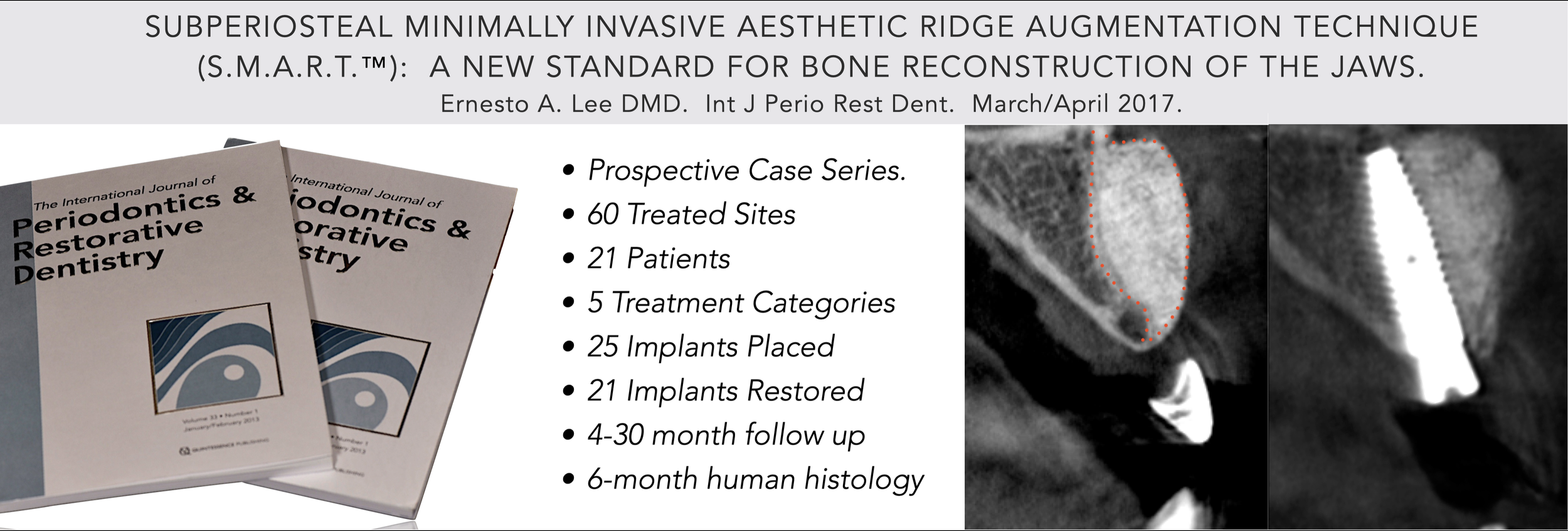
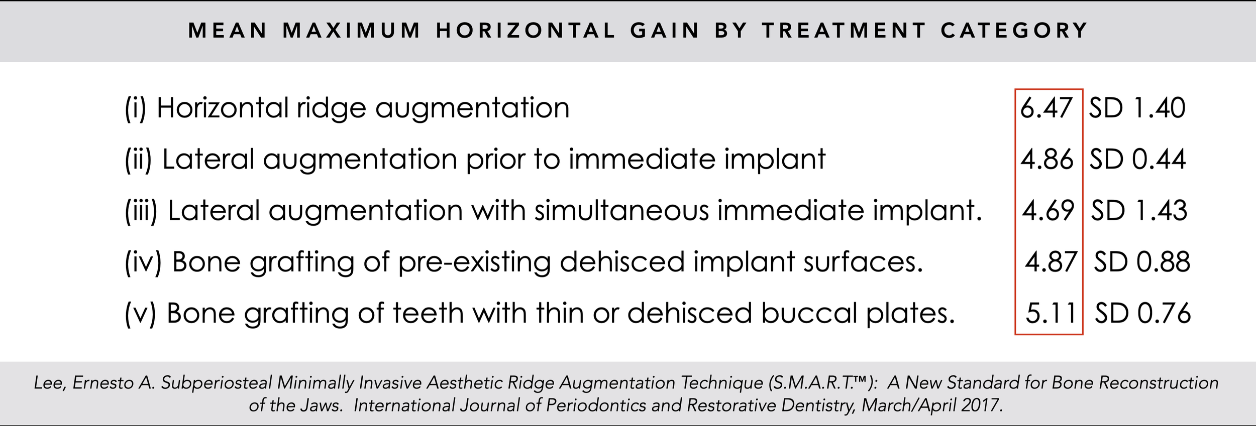
Dr. Ernesto Lee published the results of his S.M.A.R.T.® Minimally Invasive Bone Grafting research in the prestigious International Journal of Periodontics and Restorative Dentistry. This study included 60 sites, 5 clinical applications, and human histology. The results demonstrate consistently superior outcomes, with fewer complications and swelling.
Dr. Lee’s ongoing research into the wound healing phenomena associated with bone regeneration in the S.M.A.R.T.® technique culminated in the publication of a landmark article in January 2024. This study, utilizing sequential human histologic analysis, provided irrefutable evidence validating the effectiveness of the S.M.A.R.T.® method.
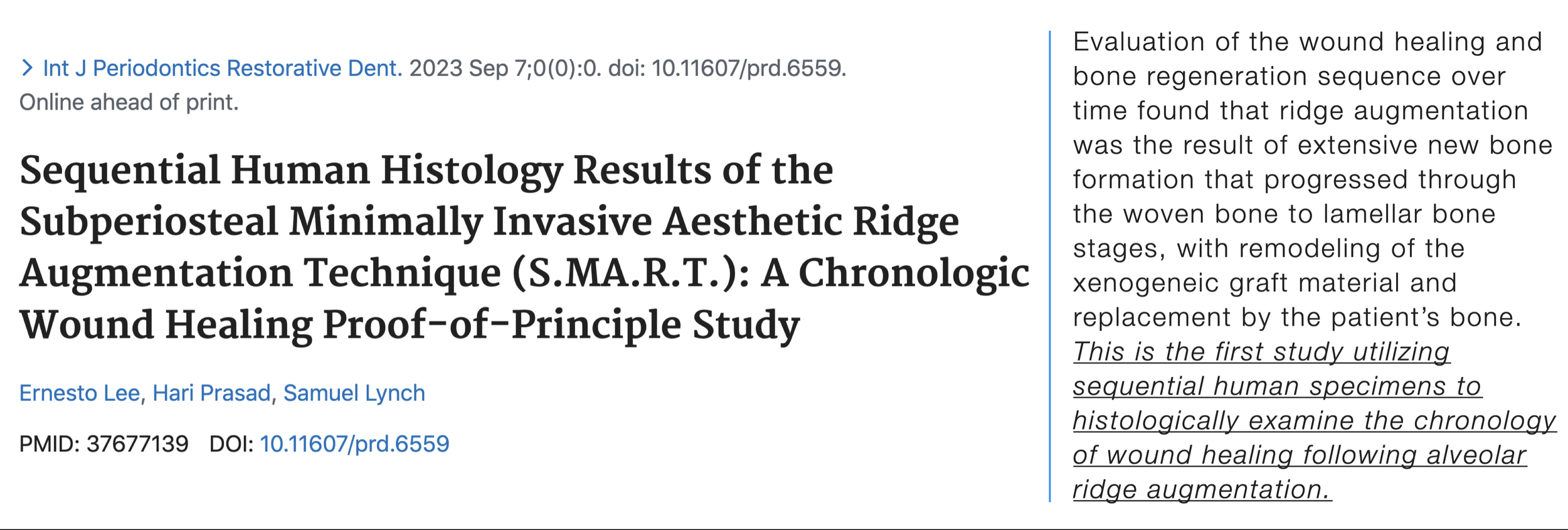
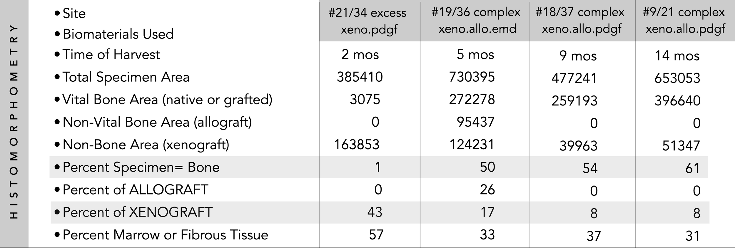
This study provides histologic proof of principle that both vertical and horizontal ridge augmentation can be successfully achieved using the S.M.A.R.T.® technique. The research documents the chronological changes in vital bone percentage, freeze-dried bone allograft (FDBA), and bovine xenograft content, as well as the formation of new bone resulting from the S.M.A.R.T.® method. The findings demonstrate that bovine xenograft resorbs more rapidly when combined with recombinant human platelet-derived growth factor-BB (rhPDGF-BB) and is subsequently replaced by regenerated bone. The reported cases highlight the technique’s ability to generate substantial bone volume for the treatment of complex three-dimensional defects—without complications—through a minimally invasive approach. Notably, this is the first study to use sequential human histologic specimens to chronologically examine bone regeneration following alveolar ridge augmentation.
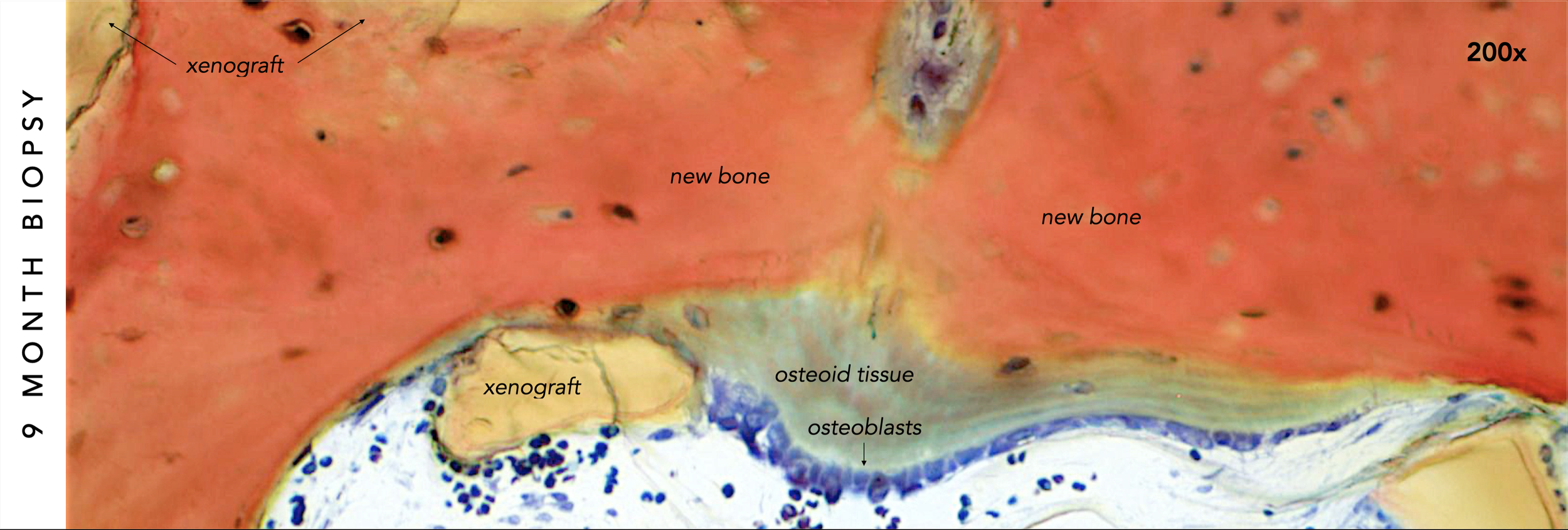
A high-magnification photomicrograph included in the article clearly depicts osteoblasts actively depositing a layer of osteoid tissue over newly regenerated bone in a 9-month histologic specimen.
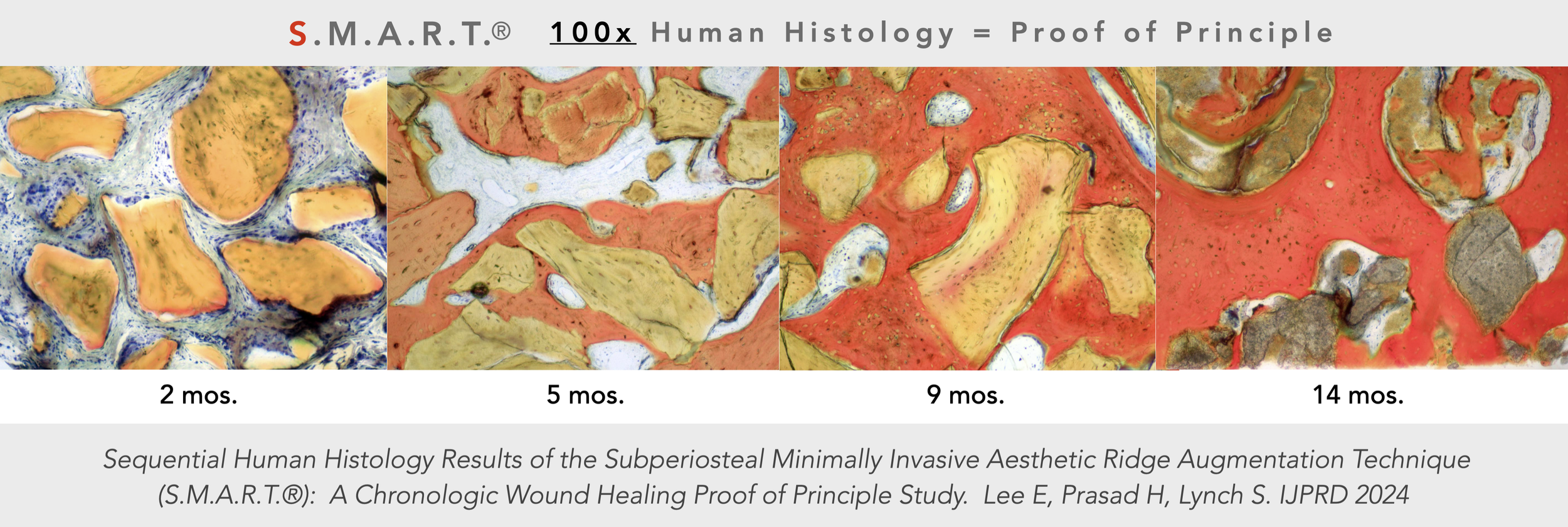
Human histologic evidence validates the bone regeneration achieved through the S.M.A.R.T.® technique, tracing its progression from early formation to mature lamellar bone. The 2-month specimen reveals a thin layer of new bone around the bovine xenograft. Over time, the xenograft particles begin to resorb, while successive remodeling cycles lead to the progressive deposition of new bone. These layers eventually connect to form bony bridges, providing structural stability to the graft. As the regenerative process continues, the new bone thickens and coalesces, creating a robust matrix that fully encapsulates the remaining xenograft particles. This dynamic sequence is clearly observable across the 5-, 9-, and 14-month specimens. Histomorphometric analysis supports these findings, demonstrating an increase in the percentage of vital bone and a corresponding decrease in xenograft content over time.
Lee E, Prasad H, Lynch S. Sequential Human Histology Results of the Subperiosteal Minimally Invasive Aesthetic Ridge Augmentation Technique (S.M.A.R.T.®): A Chronologic Wound Healing Proof-of-Principle Study. International Journal of Periodontics and Restorative Dentistry. January 2024.